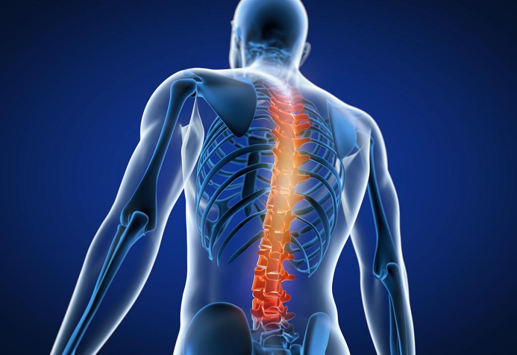
Spondylolisthesis is one of common spinal conditions that affects the lower vertebrae (spinal bones). This disease causes the lower vertebrae to slip forward onto the bone directly beneath it. It is a painful condition but treatable in most cases. Spondylolisthesis can be found in both young and elderly people, however, with different causes:
- In adolescent patients: the cause of spondylolisthesis mainly involves spondylolysis –a spine condition that results in small fracture in bones of the spine which might be a birth defect in part of the spine, causing it to slip forward during childhood or repetitive trauma to the spine. This condition is common in athletes, such as gymnasts and weightlifters.
- In elderly patients: Spondylolisthesis in the elderly is often caused due to the degenerative conditions of spine, intervertebral discs and facet joints (the joints connecting each of vertebrae). As a result, degenerative changes in these spinal structures can eventually induce spinal instability and cause vertebrae to move and slip out of place.
Causes of spondylolisthesis
Although the causes vary among individuals, the major cause of back pain in spondylolisthesis is spinal instability which is a condition that occurs when the inter-vertebral discs in the spine begin to degenerate. The bulge of the disc decreases and begins to lose height. This causes the vertebrae to displace from their anatomical position and override the disc. Once the vertebrae slip out of place, spinal canal becomes narrow, compressing the nerves traveling through the lower back into the legs, causing back pain radiating to the legs, numbness and weakness in one or both legs. If condition progresses and nerves are severely compressed, patients might experience bladder dysfunction, such as urinary retention or incontinence (loss of control). While it may affect younger patients, due to developmental causes, it is more often a degenerative condition that substantially influences elderly people.
Symptoms of spondylolisthesis
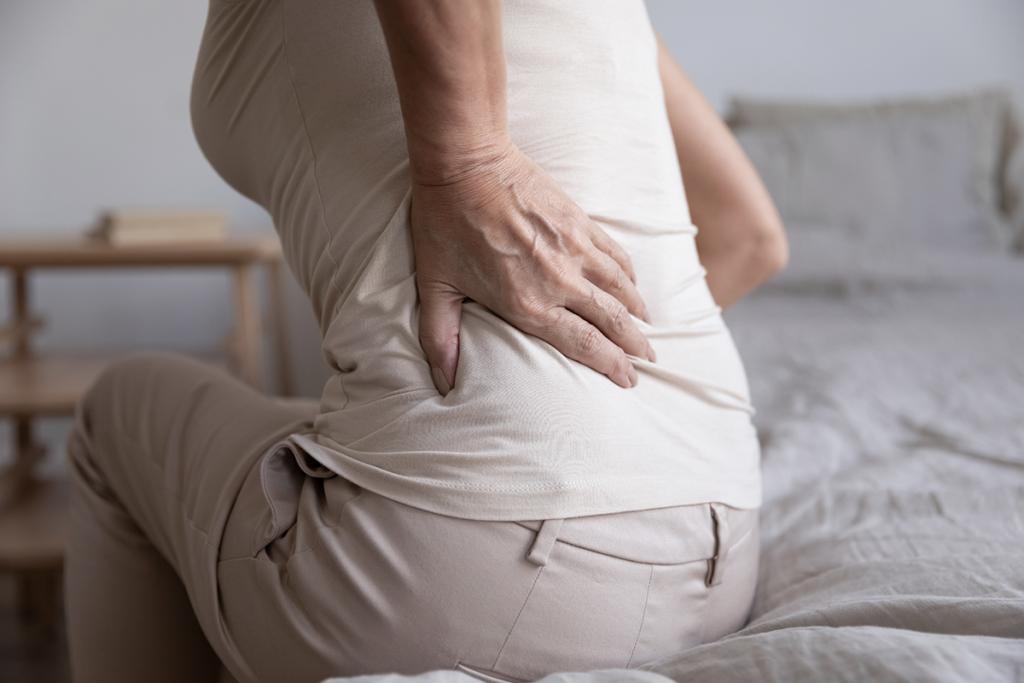
Main symptoms of spondylolisthesis usually include back pain and pain that travels from the back down one or both legs. Nevertheless, both symptoms might not be concurrently present at the same time even though most patients experience combined symptoms. In fact, symptoms can range in severity from non-existent to a loss of urination and bowel movement control in more severe cases. Pain severity widely varies. Some patients have back pain as a dominant symptom while pain radiating to the leg (s) might be a major symptom in some patients.
In asymptomatic patients, spondylolisthesis is often discovered during imaging tests. In severe cases, progressive symptoms include:
- Pain in the lower back, hip and the back of thigh (the hamstring)
- Back pain in lower back that is aggravated by back movement, twisting or bending. Pain is alleviated while lying down or resting.
- Pain that radiates to the leg (s) due to nerve compression (a pinched nerve), causing numbness in the legs or feet and leg weakness as well as urinary and fecal incontinence.
Diagnosis of spondylolisthesis
Diagnosis starts with a physical examination and imaging tests using X-ray and CT scan (Computerized Tomography). In case that nerve compression is suspected, MRI scan (Magnetic Resonance Imaging) for highly detailed images might be additionally required.
Result of X-ray scan
- X-ray might show a crack or stress fracture in the pars interarticularis which is a small segment of bone that joins the facet joints in the back of the spine.
- If the fracture gap at the pars interarticularis has widened and the vertebrae has shifted forward, it is an indication of spondylolisthesis. An x-ray taken from the side helps to determine the amount of forward slippage.
- Spinal instability is shown in flexion – extension lateral view while bending forwards and backwards.
Result of CT scan
- CT scan provides more detailed than plain x-rays.
- CT scans can identify fracture or slippage and it can be used in planning treatment if surgery is indicated.
Result of MRI scan
- MRI scan delivers a highly detailed images of soft tissues around the spinal area, including intervertebral discs, nerves, muscles and spinal ligaments.
- MRI can also determine if there is damage to the intervertebral disks between the vertebrae or if a slipped vertebra is pressing on spinal nerve roots. It can be used to guide the surgical treatment, if needed.
Treatment goals
The goals of treatment for spondylolisthesis are to:
- Alleviate significant pain
- Allow a recent pars fracture to heal (not applicable to all cases)
- Fix compressed nerves
- Realign the slipped vertebrae
Treatment of spondylolisthesis
Treatment of spondylolisthesis can be divided into 3 main approaches:
- Nonsurgical treatment
As a conservative option, nonsurgical treatment is always considered an initial treatment for most patients with low-grade spondylolisthesis. Related symptoms usually improve after receiving nonsurgical treatments which include:- Avoiding sports and other vigorous activities that place excessive stress on the lower back for a period of time can usually improve back pain and other related symptoms.
- Taking pain killer medicines e.g. paracetamol and nonsteroidal anti-inflammatory drugs (NSAIDs), such as ibuprofen, diclofenac, etoricoxib and celecoxib can help relieving back pain.
- Physical therapy. Specific exercises can help improve flexibility, stretch tight hamstring muscles and strengthen muscles in the back, hip and abdomen.
- Wearing a back brace for a period of time aims to reduce pressure on and limit movement in the lower spine.
- Spinal intervention and pain management
Spinal interventions are image-guided procedures conducted through a precisely placed, small needle aiming to provide therapeutic relief from pain associated with nerve or facet joint compression. These minimally invasive procedures are, for example, epidural injections (the injection of anesthetic and steroid medications into the epidural space to relieve pain), selective nerve root block injection and radiofrequency ablation. Not only to relieve pain, but these minimally invasive procedures also pinpoint the exact source of pain, allowing for source of pain identification. Due to high degree of safety and efficacy, spinal interventions have been widely recognized as one of standard therapeutic options for treating spine diseases. - Surgical treatment
If the patients have progressive pain which does not respond to nonsurgical treatments e.g. pain relief medications, physiotherapy and spinal interventions, surgical treatment may be recommended. Indications of spinal surgery include:- Severe or high-grade slippage or slippage that is progressively worsening
- Severe back pain that has not sufficiently improved after receiving nonsurgical treatments for a period of time
- Severe nerve compression
The goals of surgical treatment
- Decompression: aiming at relieving symptoms caused by pressure on a nerve due to a narrowed spinal canal e.g. pain radiating to the leg, numbness, weakness in the leg and urinary or fecal incontinence.
- Restabilization: focusing on correcting spinal instability and deformity that cause back pain while optimizing the physiological weight-bearing function.
- Realignment: aiming at correcting and adjusting spinal curving while improving spinal balance in patients present with scoliosis (a sideways curvature of the spine) and/or kyphosis or hunchback – a condition in which the spine in the upper back has an excessive curvature or forward rounding of the back, which leads to a slouching posture.
Surgery options
Surgeries to treat spondylolisthesis include:
- Decompression alone
Lumbar decompression surgery is spinal surgery used to treat compressed nerves in the lower spine. The surgery aims to relieve pressure and improve symptoms, such as persistent pain and numbness in the legs caused by pressure on the nerves in the vertebrae. Due to advances in surgical technology, microscopic decompression is a minimally invasive surgery performed by using microscope, resulting in smaller incisions, less pain, fewer postoperative complications and faster recovery. Since this surgery is not aimed for spinal stabilization, it might be considered only in patients who have mild to moderate pain classified as mild-grade spondylolisthesis in which the vertebrae have slightly slipped. - Spinal fusion surgery
Spinal fusion is essentially a welding process. It is conducted in order to fuse together the slipped vertebrae so that they heal into a single, solid bone. Fusion eliminates motion between the damaged vertebrae and alleviates pain caused by spinal instability. This procedure might be considered in patients who have severe back pain.
Spinal fusion techniques
There are two main types of spinal fusion surgery:
- Posterolateral Fusion
Posterior fusion is considered a conventional approach. During the procedure, the spine surgeon makes a large incision down the middle of the lower back. To access the vertebrae, the spine surgeon pulls back the muscles that surround the spine. After the decompression, the spine surgeon places graft material along the sides of the vertebrae to stimulate bone growth. The bone graft material is typically placed over the transverse processes of the vertebrae which is called a posterolateral fusion. Pedicle screws and rods are often used to provide immediate stability to the spine until a solid fusion has been achieved. These screws typically are not removed even after the bone graft has healed. Disadvantages of this techniques might involve larger incision, more pain, more blood loss and delayed recovery. In addition, the fusion rates after surgery are considerably low. - Interbody Fusion
An interbody fusion is new spinal fusion that involves removing the intervertebral disc and replacing it with a bone spacer containing bone graft material that promotes bone healing and facilitates the fusion. This type of fusion can be performed by using different approaches, depending different accesses to the intervertebral disc e.g. PLIF (Posterior Lumbar Interbody Fusion), TLIF (Transforaminal Lumbar Interbody Fusion), DLIF (Direct Lateral Interbody Fusion), ALIF (Anterior Lumbar Interbody Fusion) and OLIF (Oblique Lumbar Interbody Fusion). Since these procedures can be conducted through smaller incisions, superior advantages over conventional approach are less pain, less blood loss, faster recovery and higher infusion rates after surgery. These interbody fusion techniques have been widely accepted as standard treatments in USA and Europe; however, these approaches must be performed by well-trained and highly expert spine surgeons in conjunction with cutting-edge technology and advanced surgical equipment.
ERAS protocol for enhanced recovery after surgery
Due to surgical advances in minimally invasive spine surgery, Enhanced Recovery After Surgery (ERAS) protocol has emerged as a multimodal perioperative care pathway designed to achieve early recovery after minimally invasive spine surgery by maintaining pre-operative organ function and reducing the profound stress response following surgery while enhancing early mobilization, allowing for a quick return to daily life and activity.
At Spine Center, Bangkok International Hospital, ERAS protocol has been deployed in combination with our expertise and cutting-edge technology in order to achieve the best possible treatment outcomes with smaller incisions, less pain, less operative complications and faster recovery. ERAS protocol in spondylolisthesis surgery involves specialized care obtained from multidisciplinary approach, including these following specialties:
- Spine surgeons: If applicable, spine surgeons preferably consider “MISS” or Minimally Invasive Spine Surgery using special techniques and surgical devices through smaller incisions. Compared to conventional surgery in which large incisions are required, MISS results in less pain and less traumatic injuries to surrounding muscles and nerves, leading to faster recovery and shorter hospital stay.
- Anesthesiologists: Anesthesiologists play a significant role in delivering ERAS benefits to the patients, including minimizing pain, reducing opioid administration and undesired side effects, expediting patient recovery and enhancing early mobilization while decreasing hospital length of stay.
- Physiatrists and rehabilitation specialists: Physiatrists and rehabilitation team optimize patient’s physiologic function and facilitate recovery process. To ensure a quick recovery, physical assessment for each patient will be conducted by rehabilitation team. Pre-surgical instructions and physiotherapy advices can be given prior to surgery, resulting in faster recovery and quick return to normal tasks.
- Internal medicine physicians: Internal medicine physicians help to arrange medical plans before and after surgery particularly in patients who have had underlying conditions, such as hypertension, dyslipidemia, diabetes, cardiovascular diseases and other vascular problems.
- Nurses, pharmacists and nutritionists: Post-operative care must be also provided under close supervision of other medical professions, including nurse, clinical pharmacist and nutritionist. Medication administration and appropriate nutrition are vital for enhancing recovery process.
Spondylolisthesis is a complex condition that requires expertise from multidisciplinary team highly specialized in spine care. With our minimally invasive spine surgery conducted through ERAS protocol, patients can rest assured to receive the best possible treatment outcomes with high degree of effectiveness and safety while preserving their quality of life.